Categories
Immunotherapy Public Health

How Bacteriophages Could Save Humanity from Antibiotic Resistance

“Thanks to penicillin… he will come home!” pronounced a Life magazine advertisement published in 1944. At this time, penicillin, the first true antibiotic drug, had just been discovered and made commercially available.

Antibiotics are drugs that prevent or treat bacterial infections. Before the advent of penicillin, the leading causes for death were bacterial infections resulting in pneumonia, tuberculosis, diarrhea, and enteritis, causing one third of all deaths in the United States. Since then, the idea of deaths by bacterial infection have largely faded into the past—until the evolution of antibiotic-resistant bacteria has now threatened this status quo.

In antibiotic or antimicrobial resistance, antibiotics are no longer effective against bacteria that have evolved to survive it, particularly using beta-lactamase enzymes. This resistance is further accelerated by excessive, unnecessary use of antibiotics, mainly in industrial livestock production and over-prescription. This increased use contributes to the evolutionary pressure on microbes to develop resistance to antibiotics.

Alexander Fleming, who discovered penicillin, was receiving the Nobel Prize in medicine and physiology when he ominously predicted antibiotic resistance: “It is not difficult to make microbes resistant to penicillin in the laboratory by exposing them to concentrations not sufficient to kill them.” Fleming himself was hesitant of widespread antibiotic use, recognizing its resistant capabilities from the year they were released. In 2019, the CDC reported more than 2.8 million antibiotic-resistant infections in the US.

Antibiotics have very specific mechanisms of action to target bacteria. For example, penicillin binds to an enzyme on the bacteria and removes it, which breaks an important barrier in the cell. If one certain bacteria has a mutated enzyme, the antibiotic will be rendered ineffective, and that mutation will become prevalent in that bacterial species.

A key difference between bacteria and most other organisms is their ability to transmit genes to nearby bacteria in a process called horizontal gene transfer. This is why bacterial resistance has quickly become an epidemic as it spreads quickly and efficiently. Unfortunately, it would take almost a decade to modify the antibiotics necessary to combat these “superbug” bacteria.

Bacteriophages, meaning “bacteria eaters,” are viruses that only infect bacteria. Most are lytic, meaning that when infecting a host, they inject their genes into the host, utilize the host to rapidly replicate, and destroy the cell walls by bursting through, essentially creating a “phage-producing factory” from a bacteria. A small number are lysogenic, which means they coexist with bacteria.

Consequently, bacteriophages have provoked the interest of researchers as a potential replacement for traditional antibiotics, which are obtained from fungi. While there are a little over 100 known traditional antibiotic drugs to fight the near-infinite supply of bacteria in the world, estimates show that there exist about 10 phages for each bacterium. This indicates that there may be many more potentially therapeutic bacteriophages than traditional antibiotics. Bacteriophages are the most abundant “organism” in the biosphere, either living harmoniously with bacteria in the lysogenic cycle or destroying about 40% of the ocean’s bacteria every day, amounting to 10²³ phage infections in only one second.

Though, phages have drawbacks as potential antibiotic therapies. For one, they are extremely specific. While a single traditional antibiotic can target a multitude of bacteria, bacteriophages target one bacteria. A working solution for this is the use of “phage cocktails,” which combine multiple natural and synthetic bacteriophages to more effectively treat patients.

Also, phages are not entirely shielded from bacterial resistance. Bacteria can fight back with certain immune responses, specifically CRISPR Cas  systems. However, unlike antibiotics, phages are continually adapting and responding to such defensive systems. This continual mutation of phages poses a risk for FDA approval, but some researchers are working on machine learning  systems to predict these changes.

Still, bacteriophages show promise as an alternative therapy to traditional antibiotics. Scientists hope that phages will become an alternative defense against bacteria that could help ease antimicrobial resistance.

References
Categories
Cardiology Immunotherapy

CAR T Cells Produced by mRNA Injection Reduce Cardiac Fibrosis, Restore Function to Heart

Researchers at the University of Pennsylvania’s Perelman School of Medicine have published a method to treat cardiac fibrosis using an mRNA injection that enables an individual’s own CAR T cells to fight the disease.

Background

Cardiac fibrosis is a medical condition caused by many different types of heart disease that can lead to scarring and stiffening in the muscle wall of the heart. Normally, cells in the heart called cardiac fibroblasts help to develop the heart and maintain its homeostasis (that is, it helps the heart stay in a stable condition). However, in a patient with cardiac fibrosis, these cells no longer perform their normal function. Following a cardiac injury, fibrosis can progress from scarring to complete heart failure.

T cells are a type of white blood cell that play a key role in immune response, killing cells that they recognize to be infected with viruses, cancers, or certain other pathogens. Chimeric antigen receptor (CAR) T cells are T cells that have been engineered to recognize specific proteins as harmful. This enables them to target and kill cells that have proteins from diseases that they otherwise would not recognize as harmful.

Innovation

The Penn researchers developed a CAR T-cell therapy that works by engineering T cells to recognize and kill cells that express (create) the fibroblast activation protein (FAP), a protein key to the pathology of cardiac fibrosis. Killing FAP-expressing cells consequently treats cardiac fibrosis.

By encoding a messenger RNA (mRNA) strand that results in the creation of CAR T cells that target FAP, the researchers had the idea to deliver them to a patient’s cells through an injection containing the mRNA within a lipid nanoparticle.

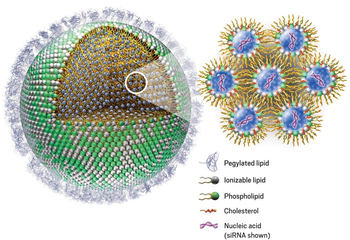
Lipid nanoparticles (LNP) are a relatively new technology discovered in the 1990s. To deliver an mRNA strand into cells to provoke a protein-expressing response, the mRNA is inserted into a sphere made of lipids that is injected into a patient. This then allows cells to uptake the LNP through endocytosis (bringing material into the cell). The mRNA then exits the LNP, causing the cell to read the mRNA instructions to create the desired protein.

09908-feature1-nanoparticle.jpg
Structure of the LNP. / Genevant Sciences via ACS.org

Without the LNP, mRNA would be unable to enter cells. mRNA vaccines for COVID-19 are a prominent use of this technology, as the mRNA that gives cells instructions to create the spike protein is protected and brought into cells by LNP.

Results

In rodents with cardiac fibrosis, the Penn researchers revealed that their mRNA injection successfully resulted in the creation of FAP-targeting CAR T cells. Observing the hearts of rodents before and after treatment showed notable improvements in cardiac function. This means that as the CAR T cells killed cells that expressed FAP, fibrosis was reduced.

Video of rodent echocardiograph recorded two weeks after treatment with CAR T-cell therapy that was given after an injury that caused cardiac fibrosis. / Rurik et al., 2022

In rodents with injuries causing cardiac fibrosis, the CAR T-cell treatment halved the percentage of fibrosis in the ventricles.

Discussion

The implications of this new treatment are of great significance. Reduction of fibrosis and restoration of cardiac function in rodents with cardiac fibrosis reveals a promising new form of treatment for human patients with the potentially fatal disease.

According to the CDC, about 659,000 people in the United States die from heart disease each year, accounting for 1 in every 4 deaths–all costing the country hundreds of billions of dollars each year. Thus, biotechnological innovations in treatment of cardiac disease can have a great impact.

Earlier CAR T-cell therapies have required a patient’s T cells to be extracted from blood, sent to a lab, engineered to find and kill certain targets, then returned intravenously to the patient. This is an extremely time-consuming and cost-prohibitive process, potentially costing patients hundreds of thousands of dollars.

The innovation of using mRNA injections to create CAR T cells within a patient’s own body instead of a lab may greatly reduce the time and financial burdens associated with CAR T-cell therapies. Rather than extracting, modifying, and replacing T cells from each patient, mRNA shots that provoke the creation of CAR T cells can be mass-produced and given to any patient.

The scope of this innovation reaches far beyond cardiac fibrosis, as it can potentially be applied to CAR T-cell therapies for cancer and other diseases.

References
Categories
Immunotherapy Oncology

Mass-Producible Specialized T Cells Exhibit High Cancer-Killing Efficacy, Minimized Complications

UCLA researchers have shown in preclinical studies that their mass-producible engineered invariant natural killer T (iNKT) cells demonstrate promising antitumor efficacy and low immunogenicity (unwanted immune response) compared to current cell-based immunotherapy for cancer treatment.

Invariant natural killer T (iNKT) cells are specialized T cells that are notable for their speedy response to danger signals and activation of macrophages (white blood cells that destroy cancer cells, microbes, cellular debris, and foreign substances).

Dr. Lili Yang’s UCLA lab generated iNKT cells by engineering hematopoietic stem cells (HSCs, precursors to all types of blood cells). These cells are allogeneic—they are not genetically specific to patients. Normally, in the realm of cell-based immunotherapy, this would be expected to cause an immune response in the form of graft-versus-host disease (GvHD), a condition in which donor stem cells attack the recipient. The study mentioned that such immunogenicity can also decrease efficacy of therapeutic cells. Therefore, allogeneic cells have not been widely used for T-cell-based therapies, with most therapies using autologous (from the patient) cells instead.

Generally, autologous T-cell therapy requires a patient’s T cells to be extracted from blood, sent to a lab, engineered to find and kill cancer cells, then returned intravenously to the patient—all costing hundreds of thousands of dollars.

Unexpectedly, when tested on mice, the Yang Engineering Immunity Lab’s allogeneic HSC-iNKT cells did not cause the negative effects associated with allogeneic cells. The researchers found that while other types of allogeneic T cells killed mice by GvHD after 2 months of cell transfer, the mice that received HSC-iNKT cells sustained long-term survival.

Figures showing (G) experimental design, (H) mouse tumor imaging, (I) quantification of tumor size based on imaging, (J) survival curves of mice over 4 months following tumor challenge.

Following irradiation of mice, those without cell therapy (labeled as vehicle) died of tumors within 45 days. Those treated with allogeneic BCAR-T cells were tumor-free but died of GvHD. Only those treated with allogeneic HSC-iNKT were tumor-free and survived long term.

This important development means that cell-based cancer therapies would no longer have to rely only on autologous cells extracted from each individual patient. Instead, with the advent of the Yang lab’s one-size-fits-all allogeneic solution, therapeutic cells could be mass-produced and given to any patient, significantly bringing down treatment costs.

The reason why allogeneic HSC-iNKT cells do not cause GvHD is currently unknown to researchers.

Graphs showing tumor load of irradiated mice over time. The Yang lab’s HSC-iNKT cells are shown to have decreased tumor load to near-zero levels (p < 0.001), a more significant decrease than was shown by the other tested therapy (PBMC-NK).
Frozen and fresh allogeneic HSC-iNKT cells were shown to kill more live lung cancer cells (H292-FG) than PBMC-NK cell therapy.

The study also showed that both frozen and fresh allogeneic HSC-iNKT cells killed live leukemia, melanoma, lung cancer, prostate cancer, and multiple myeloma cells in vitro. Compared to PBMC-NK cells, the Yang lab’s cells displayed greater tumor-killing efficacy. Importantly, allogeneic HSC-iNKT cells were also found to remain functional following freezing and thawing, which is crucial for their viability as a widespread, mass-produced treatment.

Factors that support allogeneic HSC-iNKT cells’ prospects as a future widespread cancer therapy include remaining functional following freezing and thawing, high tumor-killing efficacy, and mass-producible by virtue of low immunogenicity.

Dr. Yang told the UCLA Newsroom that one peripheral blood donation could yield 300,000 doses. The researchers are now focused on streamlining manufacturing processes, hoping to better enable mass-production, potentially bringing it to clinical and commercial development more quickly. The Newsroom noted that clinical trials have not yet occurred—this therapy has yet to be tested in humans or evaluated by the FDA. The UCLA Technology Development Group has filed a patent application for this method.

This article is based on the following sources

UCLA scientists make strides toward an ‘off-the-shelf’ immune cell therapy for cancer. (2021, November 16). UCLA Newsroom. https://newsroom.ucla.edu/releases/off-the-shelf-immune-cell-therapy-for-cancer
– Yang, L., et al. (2021, November 16). Development of allogeneic HSC-engineered iNKT cells for off-the-shelf cancer immunotherapy. Cell Reports Medicine. https://doi.org/10.1016/j.xcrm.2021.100449