Categories
Cardiology

Neural Network Outperforms Physicians at Predicting Cardiac Arrest Risk

Intro

A study published by researchers from Johns Hopkins University highlights new artificial intelligence tools that could help physicians preemptively identify cardiac arrest in patients with the use of artificial intelligence. This new technology could change the way healthcare professionals approach preventative cardiac care, potentially saving patients from fatal outcomes.

Background

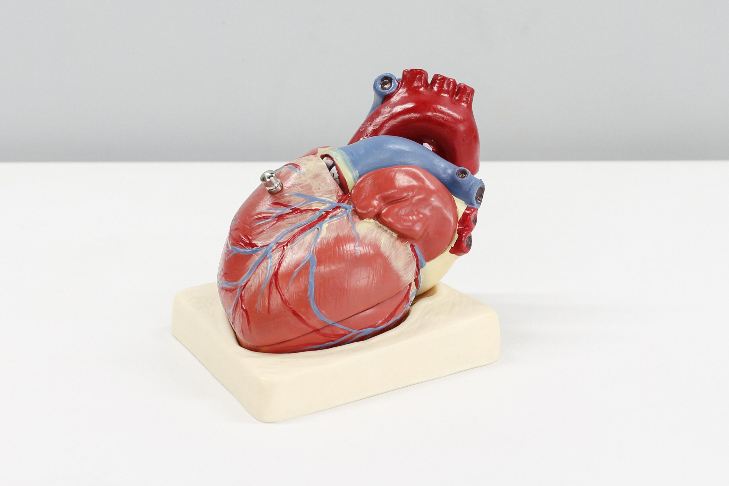
Cardiac arrest is one of the leading causes of death, causing hundreds of thousands of deaths per year in the United States. It is caused by a sudden, often arbitrary failure of the heart and can be attributed to factors such as genetics, diet, arrythmia (abnormal heartbeat), and underlying heart problems. They can be the result of chronic cardiovascular conditions but can also occur in healthy individuals. Despite the vast research regarding the disease, scientists are still not completely sure how cardiac arrest arises in patients. Cardiac arrest is nearly impossible to predict with accuracy, making it one of medicine’s deadliest and most confusing conditions.

Currently, physicians determine a patient’s likelihood of cardiac arrest by analyzing their vitals and heart scarring. Testing of vitals entails quantitative analysis of a patient’s blood, including but not limited to cholesterol and sugar levels. Heart scars are tiny marks in the heart which cause cardiovascular disease, and ultimately, cardiac arrest. However, heart scars are incredibly hard to detect because they are microscopic in size. The team of researchers from Johns Hopkins University sought to develop a solution that could accurately predict cardiac arrest risk.

Methods & Results

The team created an artificial intelligence (AI) program built on a neural network that can predict a patient’s probability of developing a cardiac arrest in the next ten years with statistically significant accuracy. The AI program views close-up images of patients’ cardiac tissues, and combined with the patient’s history, determines the probability of a cardiac arrest. The model was able to outperform human predictions of cardiac arrest, and the research team plans to implement the technology as a valuable tool available to physicians.

The research team modeled the AI after a neural network, which is a computer system modeled after the human brain. That is, “neural” pathways are strengthened by successful predictions of correlations in a given data set, enabling computers to make highly accurate predictions of increasingly complex and abstract concepts by applying its knowledge from these data sets.

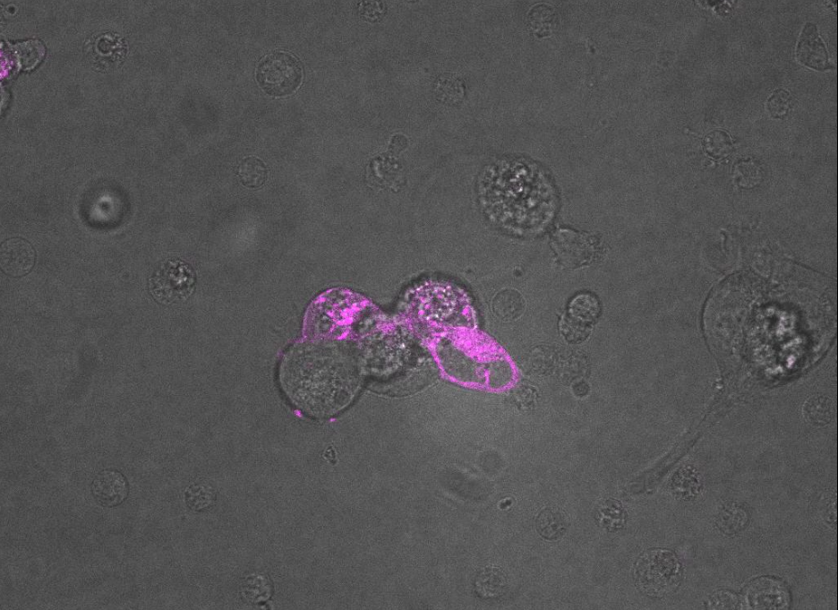
The AI was programmed to conduct a personalized, patient-specific survival assessment, which analyzes a patients’ underlying conditions and vitals. Next, the team used contrast-enhanced cardiac scar images from and taught the AI to detect aspects of the image that are invisible to the naked eye by using neural network technology. Currently, cardiologists are only able to analyze parts of scar images such as volume, mass, shape, etc. These enhanced images are evaluated by the AI in quantitative ways that human doctors could simply never achieve. The AI was then tested on real patients and data from previous years to see if the neural network could use this data to reliably extrapolate it onto new data.

The researchers found that their algorithm could accurately predict cardiac arrest in real patients to a better extent than physicians. They also tested the AI at 60 different health centers around the US, indicating that this model could be replicated at other hospitals.

Discussion

The researchers concluded that the AI could be of major use to physicians. They plan to continue development of the program for both cardiac arrest and other heart-related diseases. The technology could also improve the accuracy of other diagnostics that rely solely on visual observation. These findings have grand implications on the future of healthcare, indicating a new role of specialized software and artificial intelligence. It may not be long before this novel application of artificial intelligence becomes widespread among physicians, enabling improved patient care by revealing the previously unnoticed.

References
Categories
Cardiology Immunotherapy

CAR T Cells Produced by mRNA Injection Reduce Cardiac Fibrosis, Restore Function to Heart

Researchers at the University of Pennsylvania’s Perelman School of Medicine have published a method to treat cardiac fibrosis using an mRNA injection that enables an individual’s own CAR T cells to fight the disease.

Background

Cardiac fibrosis is a medical condition caused by many different types of heart disease that can lead to scarring and stiffening in the muscle wall of the heart. Normally, cells in the heart called cardiac fibroblasts help to develop the heart and maintain its homeostasis (that is, it helps the heart stay in a stable condition). However, in a patient with cardiac fibrosis, these cells no longer perform their normal function. Following a cardiac injury, fibrosis can progress from scarring to complete heart failure.

T cells are a type of white blood cell that play a key role in immune response, killing cells that they recognize to be infected with viruses, cancers, or certain other pathogens. Chimeric antigen receptor (CAR) T cells are T cells that have been engineered to recognize specific proteins as harmful. This enables them to target and kill cells that have proteins from diseases that they otherwise would not recognize as harmful.

Innovation

The Penn researchers developed a CAR T-cell therapy that works by engineering T cells to recognize and kill cells that express (create) the fibroblast activation protein (FAP), a protein key to the pathology of cardiac fibrosis. Killing FAP-expressing cells consequently treats cardiac fibrosis.

By encoding a messenger RNA (mRNA) strand that results in the creation of CAR T cells that target FAP, the researchers had the idea to deliver them to a patient’s cells through an injection containing the mRNA within a lipid nanoparticle.

Lipid nanoparticles (LNP) are a relatively new technology discovered in the 1990s. To deliver an mRNA strand into cells to provoke a protein-expressing response, the mRNA is inserted into a sphere made of lipids that is injected into a patient. This then allows cells to uptake the LNP through endocytosis (bringing material into the cell). The mRNA then exits the LNP, causing the cell to read the mRNA instructions to create the desired protein.

09908-feature1-nanoparticle.jpg
Structure of the LNP. / Genevant Sciences via ACS.org

Without the LNP, mRNA would be unable to enter cells. mRNA vaccines for COVID-19 are a prominent use of this technology, as the mRNA that gives cells instructions to create the spike protein is protected and brought into cells by LNP.

Results

In rodents with cardiac fibrosis, the Penn researchers revealed that their mRNA injection successfully resulted in the creation of FAP-targeting CAR T cells. Observing the hearts of rodents before and after treatment showed notable improvements in cardiac function. This means that as the CAR T cells killed cells that expressed FAP, fibrosis was reduced.

Video of rodent echocardiograph recorded two weeks after treatment with CAR T-cell therapy that was given after an injury that caused cardiac fibrosis. / Rurik et al., 2022

In rodents with injuries causing cardiac fibrosis, the CAR T-cell treatment halved the percentage of fibrosis in the ventricles.

Discussion

The implications of this new treatment are of great significance. Reduction of fibrosis and restoration of cardiac function in rodents with cardiac fibrosis reveals a promising new form of treatment for human patients with the potentially fatal disease.

According to the CDC, about 659,000 people in the United States die from heart disease each year, accounting for 1 in every 4 deaths–all costing the country hundreds of billions of dollars each year. Thus, biotechnological innovations in treatment of cardiac disease can have a great impact.

Earlier CAR T-cell therapies have required a patient’s T cells to be extracted from blood, sent to a lab, engineered to find and kill certain targets, then returned intravenously to the patient. This is an extremely time-consuming and cost-prohibitive process, potentially costing patients hundreds of thousands of dollars.

The innovation of using mRNA injections to create CAR T cells within a patient’s own body instead of a lab may greatly reduce the time and financial burdens associated with CAR T-cell therapies. Rather than extracting, modifying, and replacing T cells from each patient, mRNA shots that provoke the creation of CAR T cells can be mass-produced and given to any patient.

The scope of this innovation reaches far beyond cardiac fibrosis, as it can potentially be applied to CAR T-cell therapies for cancer and other diseases.

References
Categories
Cardiology COVID-19

American Heart Association Quells Vaccine Myocarditis Fears Amid Growing Public Concern

As concerns regarding mRNA vaccine-caused myocarditis skyrocket on social media and news outlets, researchers have published a study in the American Heart Association’s Circulation journal with statistics regarding the potential side effect.

Google Search interest in “COVID vaccine myocarditis” from December 6, 2020 to December 6, 2021. / trends.google.com

Myocarditis is a condition that causes inflammation in the heart, which can weaken its ability to regularly pump blood throughout the body. It can lead to heart failure, abnormal heartbeat, and sudden death. Most cases of myocarditis are caused by viruses, but the rare heart condition has been noticed as a potential side effect of mRNA vaccines for COVID-19.

The study published in Circulation statistically analyzed cases in patients younger than 21 years old who had received an mRNA vaccine within 30 days of showing symptoms of myocarditis. Researchers found that, in general, young people who experience myocarditis as a side effect of the COVID-19 vaccine recover quickly and completely.

The Circulation study found that 90.6% of adolescent and young adult patients who experienced myocarditis after vaccination were male. In most cases, symptoms presented 2 days after vaccination. The most common symptom was chest pain, which presented in 99.3% of the patients. 18.7% of the patients had low left ventricular ejection fraction (LVEF), meaning that not enough blood was pumping out of their hearts. However, all patients with low LVEF who followed up had fully recovered with normalized function.

Another study published in the New England Journal of Medicine reported that only 2.13 in 100,000 people who received the mRNA vaccine experienced myocarditis. This is much lower than the 150 in 100,000 rate of myocarditis in unvaccinated patients infected with COVID-19 as reported by the Morbidity and Mortality Weekly Report. This means that unvaccinated people infected with COVID-19 had about 70 times greater incidence of myocarditis than any person receiving the mRNA vaccine.

The Circulation study’s first author, Dr. Dongngan T. Truong, told the American Heart Association Newsroom that the data showed that “most cases of suspected COVID-19 vaccine-related myocarditis in people younger than 21 are mild and resolve quickly.”

As the data shows that myocarditis as a side effect of COVID-19 vaccination is extremely rare (2.13 in 100,000) and that almost all of those patients recovered quickly and completely, the American Heart Association continues holding its position that COVID-19 vaccines are safe and highly effective (preventing hospitalization and death in 91% of severe infections). Dr. Donald M. Lloyd-Jones, president of the AHA, said that COVID-19 vaccines were “fundamental to saving lives, protecting our families and communities against COVID-19, and ending the pandemic,” then urging parents to vaccinate their children as soon as possible.

References

Note from the Editors

This article does not offer medical advice. It is a review of statements and data offered by the American Heart Association. Consult with a doctor regarding concerns related to health effects from the COVID-19 vaccine.

Categories
Cardiology

Surgeons at Duke University First to Implant New Total Artificial Heart Into Patient

A team of surgeons at Duke University led by Dr. Carmelo Milano and Dr. Jacob Schroder were the first to implant a new generation of Total Artificial Heart (TAH) in a 39-year-old male patient with heart failure after receiving FDA approval for human trials.

Home - Carmat :Carmat
The total artificial heart made by French company CARMAT acts to replace that of an organic diseased heart of patients with heart failure, hoping to one day replace the need for living donor heart transplantation. (carmatsa.com)

CARMAT’s Total Artifical Heart product, called Aeson, is a new TAH that solves many of the issues with current treatment options associated with biventricular heart failure. Current options include human organ transplantation, which carries high risk of rejection. Such rejection can lead to the need for repeat cardiac transplant or even death. Other treatment options such as biventricular assist devices (BiVADs) carry high risk of neurological complication incidence such as stroke caused by accumulation of clots or seizures that can lead to life debilitating changes.

CARMAT’s Aeson is only the second TAH on the market. Aeson has made notable improvements over competitor SynCardia’s total artificial heart. CARMAT’s TAH is quieter and has a variable heart rate that adjusts based on patient activity, while SynCardia is notably louder and has a fixed heart rate.

Aeson replaces the patient’s heart by pumping blood to the pulmonary tract and systemic system. It does this through the use of battery-powered electrohydraulic rotary pumps with attached sensors that respond to changes in pressure and cardiac demands.

The French company’s device can be used as an intermediate heart prior to transplantation, or even as a complete replacement for living donor hearts. This development has radical implications to heart failure treatment, as American patients can expect to wait more than six months for a transplant.

Dr. Jacob Schroder (Assistant Professor of Surgery) and Dr. Carmelo (Professor of Surgery) installing the CARMAT TAH. (CBS17)

The surgery at Duke University was the first of its kind in the United States. It consisted of an 8-hour surgery in which Dr. Jacob Schroder and Dr. Carmelo Milano worked to remove Matthew Moore’s (a 39-year-old patient with biventricular heart failure on the transplant waiting list) left and right ventricles of his heart. They then installed the device successfully replacing the structures removed.

Each device costs around $190,000, not including the costs of critical care staff and other medications. However, this price point is significantly lower than the average cost for a human heart transplant, which is about $1.4 million.

CARMAT hopes to solve long wait lists and high costs with its TAH, Aeson, potentially offering a permanent solution in which a heart will always be available for any patient in need.

This article is based on the following sources

– Bailey, S. (2021, March 25). This new artificial heart responds to the patient. CNN Business. https://www.cnn.com/2021/03/25/business/carmat-artificial-heart-spc-intl/index.html
– Duke University School of Medicine. (2021, July 16). New generation artificial heart implanted in patient at Duke – First in U.Shttps://medschool.duke.edu/blog/new-generation-artificial-heart-implanted-patient-duke-first-us
– Rapp, N., & Vandermey, A. (2017, September 14). Here’s what every organ in the body would cost to transplant. Fortune. https://fortune.com/2017/09/14/organ-transplant-cost/
– Tan, K. (2021, July 29). Duke surgical team successfully implants new generation artificial heart in patient, first in U.S. Duke Chronicle. https://www.dukechronicle.com/article/2021/07/duke-university-hospital-health-artificial-heart-transplant-research-study-carmat
– University of California, San Francisco Health. (n.d.). FAQ: Heart transplanthttps://www.ucsfhealth.org/education/faq-heart-transplant