Categories
COVID-19

CRISPR Test Detects All Variants of COVID-19, Could Run on Mobile Phones

During the pandemic, laboratories across the world worked hard to improve current diagnostic testing methods. The main method, quantitative polymerase chain reaction (qPCR) turns a small quantity of DNA into a larger amount and uses fluorescent dyes to indicate the presence or absence of viral genetic material. However, this method falls short in the following ways:

  1. It requires expensive equipment and reagents, along with trained personnel.
  2. It requires temperature cycles, so it cannot be performed at a single temperature.
  3. It takes time. Depending on the initial amount of target present, a qPCR test can take as long as 90 minutes.

At the University of Florida, PhD student Long T. Nguyen, working under Dr. Piyush Jain, has developed a rapid, single temperature COVID-19 diagnostic test that provides results in under 30 minutes. Amazingly, the test distinguishes between five COVID variants, achieves amplification, and RNA to DNA conversion all in one “pot.” Finally, the results can be read on a mobile phone.

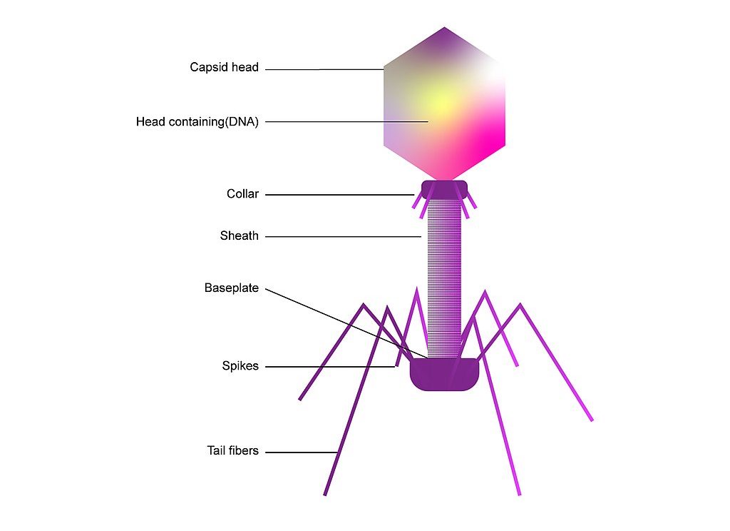
The system they used is based on a detection system found in bacteria. Bacteria contain natural immune systems called CRISPR Cas, which function to create both a memory of past viral infections, along with a defense system once these viruses come back. Cas is a protein which is sometimes described as “a pair of molecular scissors,” capable of cutting DNA or RNA fragments, while CRISPR contains complementary sequences to attacking viruses and acts as a “molecular GPS,” helping Cas find a certain target. For this reason, it is also called a Guide RNA.

DNA sequence matching the guide RNA and being cut by a Cas protein into two slices. / Javier Zarracina via vox.com

Some Cas proteins locate their target and only make cuts around the target DNA/RNA; this is called cis cleavage. Others go on a “cutting frenzy.” After finding the target and cutting, the Cas protein starts cutting up other DNA or RNA fragments surrounding it, termed trans cleavage. Cas9 proteins, famous for genetic engineering, employ cis cleavage and only cut DNA. Cas12 and Cas13 proteins utilize trans cleavage, cutting DNA and RNA respectively. All bacteria have adapted their own systems, with slight variations, allowing scientists to harness each’s individual powers.

Cas12 and Cas13 proteins are at the forefront of diagnostic research. Their cutting frenzy may not be great for gene editing, but recent innovation has found that FQ reporters, or fluorescent quenchers, can be used to detect a signal with light. These reporters are dampened by a piece of RNA or DNA located between the fluorescent and quencher. Once cut, they glow and show a light on a fluorescent reader. If a Cas12 or Cas13 protein detects its target, it will cut the target then rapidly start cutting the FQ reporters nearby.

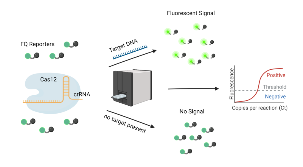
CRISPR RNA, shortened as crRNA, can be “programmed” to target any part of a target sequence. Specifically for the virus that causes COVID-19, there is a highly conserved region called the N gene. Since this same sequence is found across all variants, it can indicate the presence of the virus, but does not distinguish between mutated strains. The Jain lab identified mutated regions on each of the five variants: Alpha, Beta, Gamma, Delta, and Omicron, and created crRNAs which were complementary to each of these mutated regions. 

The N gene encodes for the nucleocapsid region on the COVID-19 virus. It is found in all variants of COVID-19.(Kubina & Dziedzic, 2020)

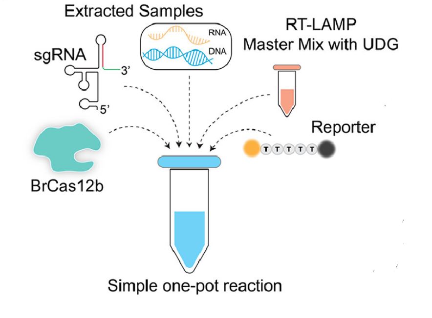
They then had to choose the optimal Cas protein, which could withstand higher temperatures. This was necessary because amplification occurs at high temperatures, ranging from 55-70°C. BrCas12b comes from a thermophilic bacterium found in hot springs and was the optimal choice.

They combined this Cas protein, along with a crRNA and finally, a master mix of RT-LAMP (Reverse Transcription Loop Mediated Isothermal Amplification). This is a very complex sounding term, but it can be broken down fairly easily. Reverse transcription is the process of converting RNA into DNA. Isothermal means it works at a single temperature and amplification implies the amount of DNA increases greatly. This amplification also provides a checkpoint. Researchers were able to first see if the patient sample was amplified; if so, COVID must be present. Then, using the CRISPR Cas system, they can determine exactly what variant is present. 

Image showing all of the components used in the Jain lab’s one-pot detection. (Nguyen et al., 2022)

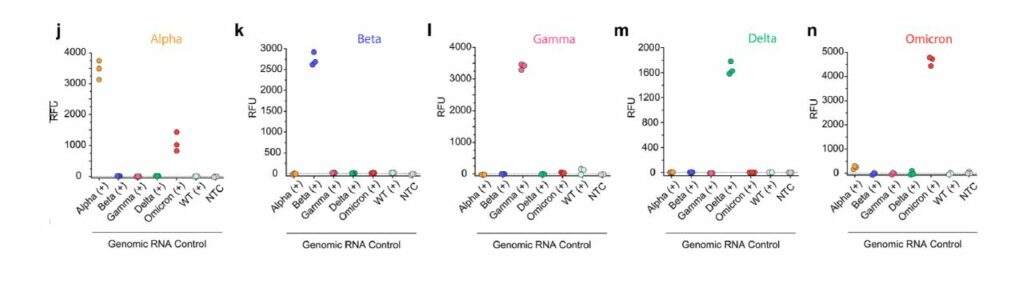
Detection is completed in under 30 minutes and patient samples with a higher viral load (i.e., they had more SARS-CoV-2 virions present in their sample), exhibited 100% accuracy with about 95% sensitivity in distinguishing variants. The figure below, from Long et. al shows the incredible accuracy which comes from this detection. The colored titles, “Alpha, Beta, etc.” are the variant present in the patient sample, while the x-axis shows the crRNA used. For instance, it was expected that if a patient contained the Beta strain, only the Beta crRNA would show high signal.

Detection results for variants, showing high sensitivity. (Nguyen et al., 2022)

Most studies combining RT-LAMP with a CRISPR reaction have extremely low sensitivity and difficulty distinguishing a positive sample. The use of specifically BrCas12b, a less studied Cas protein, allowed the Jain lab to circumvent many of the problems others have had combining the two. The applicability of this research extends far beyond COVID-19 detection. Any RNA or DNA detection could be done utilizing this research, simply by changing the sequence located on the crRNA.

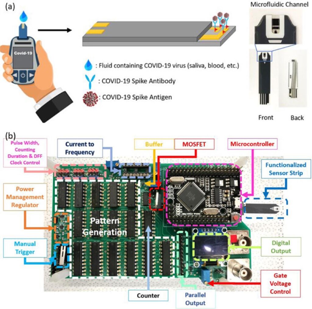
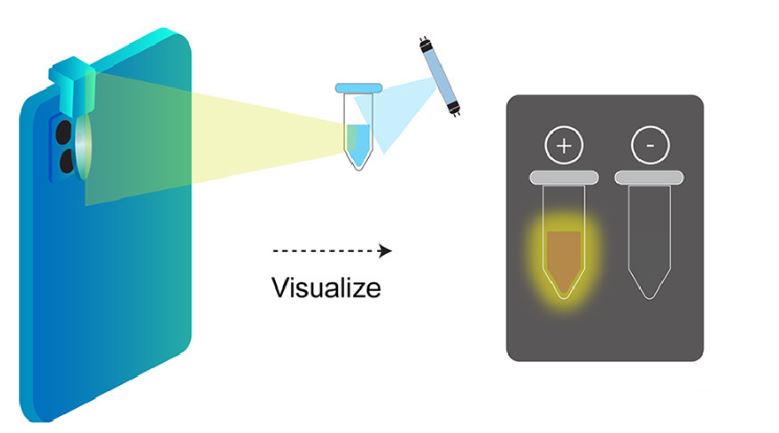
Moreover, the Jain lab aims to create portable and cheap methods for testing. They proposed an inexpensive lens which can be attached to any mobile phone camera. In a dark setting, the lens, which costs less than $5 can shine light of a specific wavelength on a sample with the added CRISPR Cas reagent and glow in the presence of COVID-19.

Potential for at-home testing using a specialized lens. Positive samples will glow in the dark once subjected to the light. (Nguyen et al., 2022)
References
  • Kubina, R., & Dziedzic, A. (2020, June 26). Molecular and serological tests for COVID-19. A comparative review of SARS-Cov-2 coronavirus laboratory and point-of-care diagnostics. MDPI. https://doi.org/10.3390/diagnostics10060434
  • Nguyen, L. T., et al. (2022, March 1). A Thermostable Cas12b from Brevibacillus Leverages One-pot Detection of SARS-CoV-2 Variants of Concern. eBioMedicine, The Lancet Discovery Science. https://doi.org/10.1016/j.ebiom.2022.103926
Categories
Immunotherapy Public Health

How Bacteriophages Could Save Humanity from Antibiotic Resistance

“Thanks to penicillin… he will come home!” pronounced a Life magazine advertisement published in 1944. At this time, penicillin, the first true antibiotic drug, had just been discovered and made commercially available.

Antibiotics are drugs that prevent or treat bacterial infections. Before the advent of penicillin, the leading causes for death were bacterial infections resulting in pneumonia, tuberculosis, diarrhea, and enteritis, causing one third of all deaths in the United States. Since then, the idea of deaths by bacterial infection have largely faded into the past—until the evolution of antibiotic-resistant bacteria has now threatened this status quo.

In antibiotic or antimicrobial resistance, antibiotics are no longer effective against bacteria that have evolved to survive it, particularly using beta-lactamase enzymes. This resistance is further accelerated by excessive, unnecessary use of antibiotics, mainly in industrial livestock production and over-prescription. This increased use contributes to the evolutionary pressure on microbes to develop resistance to antibiotics.

Alexander Fleming, who discovered penicillin, was receiving the Nobel Prize in medicine and physiology when he ominously predicted antibiotic resistance: “It is not difficult to make microbes resistant to penicillin in the laboratory by exposing them to concentrations not sufficient to kill them.” Fleming himself was hesitant of widespread antibiotic use, recognizing its resistant capabilities from the year they were released. In 2019, the CDC reported more than 2.8 million antibiotic-resistant infections in the US.

Antibiotics have very specific mechanisms of action to target bacteria. For example, penicillin binds to an enzyme on the bacteria and removes it, which breaks an important barrier in the cell. If one certain bacteria has a mutated enzyme, the antibiotic will be rendered ineffective, and that mutation will become prevalent in that bacterial species.

A key difference between bacteria and most other organisms is their ability to transmit genes to nearby bacteria in a process called horizontal gene transfer. This is why bacterial resistance has quickly become an epidemic as it spreads quickly and efficiently. Unfortunately, it would take almost a decade to modify the antibiotics necessary to combat these “superbug” bacteria.

Bacteriophages, meaning “bacteria eaters,” are viruses that only infect bacteria. Most are lytic, meaning that when infecting a host, they inject their genes into the host, utilize the host to rapidly replicate, and destroy the cell walls by bursting through, essentially creating a “phage-producing factory” from a bacteria. A small number are lysogenic, which means they coexist with bacteria.

Consequently, bacteriophages have provoked the interest of researchers as a potential replacement for traditional antibiotics, which are obtained from fungi. While there are a little over 100 known traditional antibiotic drugs to fight the near-infinite supply of bacteria in the world, estimates show that there exist about 10 phages for each bacterium. This indicates that there may be many more potentially therapeutic bacteriophages than traditional antibiotics. Bacteriophages are the most abundant “organism” in the biosphere, either living harmoniously with bacteria in the lysogenic cycle or destroying about 40% of the ocean’s bacteria every day, amounting to 10²³ phage infections in only one second.

Though, phages have drawbacks as potential antibiotic therapies. For one, they are extremely specific. While a single traditional antibiotic can target a multitude of bacteria, bacteriophages target one bacteria. A working solution for this is the use of “phage cocktails,” which combine multiple natural and synthetic bacteriophages to more effectively treat patients.

Also, phages are not entirely shielded from bacterial resistance. Bacteria can fight back with certain immune responses, specifically CRISPR Cas  systems. However, unlike antibiotics, phages are continually adapting and responding to such defensive systems. This continual mutation of phages poses a risk for FDA approval, but some researchers are working on machine learning  systems to predict these changes.

Still, bacteriophages show promise as an alternative therapy to traditional antibiotics. Scientists hope that phages will become an alternative defense against bacteria that could help ease antimicrobial resistance.

References
Categories
COVID-19

Researchers Reveal Portable COVID Testing Method, Gives Results Within One Second

Researchers from the University of Florida, along with collaborators from the National Chiao Tung University, recently created the world’s fastest COVID detection test to date using a new method with antibody-infused test strips and a small circuit board.

Since the beginning of the COVID-19 pandemic, RT-PCR tests, commonly referred to as PCR, have been regarded as the gold standard for COVID-19 testing.

Reverse Transcription Polymerase Chain Reaction (RT-PCR) works by first converting RNA into DNA, followed by copying small segments of this DNA over and over, primarily using temperature to denature and bind DNA, along with “primers” to make new copies. The process takes about two hours and uses expensive machinery. Such amplification of DNA makes it easy for machines to detect the small amounts of viral particles present in infected patient samples, but difficult to apply to large populations during a pandemic.

One of the defining features of the coronavirus is the spike proteins, which enable the virus to penetrate host cells due to their geometry and location. Rather than having to convert RNA to DNA, copy the DNA, and read a signal as is done with RT-PCR tests, a new study described a system which uses the spike protein-antibody bond and circuitry for detection.

Antibodies are Y-shaped proteins our immune system produces to fight and prevent future infection. They work by creating sites to which infectious particles bind, effectively blocking those particles from infecting cells. These sites can include binding locations for viruses such as SARS-CoV-2, which researchers have found to be quite useful for detection.


As our need for fast, cheap, and portable detection grows, researchers have been searching for new methods. The researchers from the University of Florida ingeniously combined knowledge of antibodies and circuitry to detect presence of COVID in one second.

First, they modeled commercially available glucose testing strips commonly used for testing blood sugar levels in diabetic patients. If you were to dissect a glucose test strip, you would find several electrodes, coated and made of different materials.

Most commonly, glucose test strips are coated with an enzyme that reacts with glucose to steal its available electrons. These electrons are then transported to the electrode which can detect and quantify their presence, indicating how much glucose was in the blood sample.

In the study, researchers worked to transform the electrodes using different biological and chemical materials. One of the electrodes was plated with gold then “biofunctionalized” with coronavirus antibodies.  An electrode in the middle was connected to an electronic component called a metal-oxide-semiconductor field-effect transistor (MOSFET), which is used to control and amplify electrical signals.

When spike proteins from a sample interact with the surface, the antibody-antigen complex will spring up and down, causing an electrical signal to be sent to the gate of the MOSFET. The device’s circuit board can then quickly convert and read the signal. 

The MOSFET is especially important as it can convert electrical activity from the interaction of a very small amount of coronavirus with the antibodies into a very large signal, similar to how RT-PCR tests amplify the small amount of genetic material into a much larger and easier-to-detect sample.

The accuracy and acute sensitivity of this method are a direct result of combining electrical and biological tools of detection. Not only does this allow for the detection of extremely low quantities of virus particles, but it can be accomplished in merely 1 second. Furthermore, the device is inexpensive and portable, paving the way for fast, economical, and highly sensitive at-home diagnostic kits.

Notably, Minghan Xian, first author of the study, remarked that by simply altering the type of antibody used, this detection kit could be reapplied to a multitude of other infectious diseases. The electronic components can also be reused with new electrodes.

References